Advanced Microscope Facility

Microscope facility offers basic and advanced training and access to several microscopes including Nikon A1Rsi (with PicoQuant additional module) and Leica Stellaris 8 confocal systems, Zeiss Lightsheet 7, Zeiss Elyra 7 with lattice SIM and other fluorescence microscopes and stereomicroscopes, as well as to image analysis software like Bitplane Imaris, Scientific Volume Imaging (Huygens), Nikon NIS Elements, Zeiss ZEN and Leica LAS X.
Responsible staff

Anna Kasprowicz-Maluśki, PhD
e-mail: akas@amu.edu.pl
phone: +48 61 829 5968
Wojciech Kwiatkowski, MSc
e-mail: w.kwiatkowski@amu.edu.pl
phone: +48 61 829 5971
Microscopes
NIKON A1Rsi + PicoQuant LSM
Point scanning confocal microscope with hybrid-scanner (galvano/resonant), 4 channel detection (excitation wavelengths 405, 457, 488, 514, 561 and 638 nm) and 32 channel spectral detector (up to 2,5 nm spectral resolution). Based on inverted, motorized Nikon Eclipse TiE. Fully encoded scanning XY motorized stage for multipoint time lapse and tile scanning. Piezo-Z stage insert for high-speed Z-stack acquisition (100 μm/s). Nikon Perfect Focus System for maintaining focal plane during time-lapse experiments. Dual scanner: resonant and galvano: a glavano scanner for low-noise images ranging in resolution from 64 x 64 up to 4096 x 4096 pixels and a resonant scanner that is capable of taking up to 30 full 512 x 512 pixel frames per second. Life cell imaging of mammalian cells is possible – the system is equipped with an on-stage incubation chamber, controlling temperature, CO2-concentration and humidity (okolab). The A1R can also be used together with a FLIM system from PicoQuant, consisting of pulsed lasers (440 and 485 nm) and a time-correlated single photon counting unit.

LEICA Stellaris 8
Point scanning confocal microscope with fast scanner (FOV 22 mm, scan speed up to 10 pictures / sec in 512×512 mode). Spectral detection – 4 spectral detectors built-in (3x Power HyD S detector, 1x Power HyD X detector). Based on inverted, motorized Leica DMi8. Fully encoded scanning XY motorized stage (by Marzhauser) for multipoint time lapse and tile scanning. Leica AFC system with Closed Loop Focus for maintaining focal plane during time-lapse experiments. Confocal scanner: resolution from up to 8192 x 8192 pixels that is capable of taking up to 10 full 512 x 512 pixel frames per second. Life cell imaging of mammalian cells is possible – the system is equipped with an on stage incubation chamber, controlling temperature, CO2-concentration and humidity (okolab). FAst Lifetime CONtrast (FALCON) module – for FLIM, FCS, FCCS, FLCS experiments. TauSense –application-oriented imaging tools based on fluorescence lifetime (TauContrast, TauGating, TauSeparation, TauInteraction). Lightning module – for sub superresolution imaging (up to 120 nm XY, 200 nm Z). Navigator module available. Automatic water Micro Dispenser available.

Zeiss Lightsheet 7
Light sheet fluorescence microscopy (LSFM) splits fluorescence excitation and detection into two separate light paths, with the axis of illumination perpendicular to the detection axis. That means you can illuminate a single thin section of the sample at one time, generating an inherent optical section by exciting only fluorescence from the in-focus plane. No pinhole or image processing is required. Light from the in-focus plane is collected on the pixels of a camera, rather than pixel by pixel as, for example, in confocal or other laser scanning microscopes. Parallelization of the image collection on a camera-based detector lets you collect images faster and with less excitation light than you would with many other microscope techniques. This makes 3D imaging extremely fast and very light efficient. The de-coupling of the detection optics from the illumination optics enables fluorescence excitation with dedicated lenses at low numerical aperture, without sacrificing detection resolution and sensitivity. This makes LSFM ideal for imaging of samples at the millimeter scale, such as developing organisms or large cleared tissue samples. The system is equipped with a chamber with temperature control and CO2. An additional server for data processing is connected to the system (the system generates a large volume of files).

ZEISS Elyra 7 with Lattice SIM
The Elyra 7 is a motorized inverted microscope with resolution beyond the Abbe diffraction limit of the conventional light microscope. The microscope is equipped with a super-resolution system that uses a specialized lattice illumination pattern for 3D structured illumination (SIM) images and single-molecule localization methods (SMLM) like
STORM and PALM. SIM is a technology that can be used with standard fluorophores, making it an easily accessible choice for super-resolution results. The lattice illumination allows for faster, gentler, and deeper imaging of live samples, and can reach up to 255 FPS during time-lapse acquisition. The unit has optional, external incubation chamber for live imaging with controlled CO2 and temperature. For fast and precise Z-movement there is a piezo Z stage insert.
The system yields up to 8-fold improvement in voxel resolution in the X, Y, and Z dimensions. The microscope is equipped with four solid-state lasers (405, 488, 561 and 642 nm). The signal detection is assured by 2 sCMOS cameras which enable simultaneous detection of two different fluorophores. Filter set FLEX is equipped with dual and triple emission bands and allows for fast sequential two channel acquisition by fast switching of laser lines via AOTF. The high-tech offline PC workstation with high-capacity storage assures the fast processing of acquired samples.

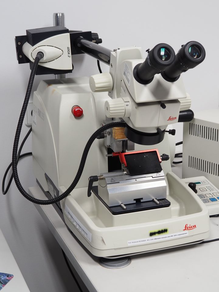
Leica RM 2255 automated rotary microtome + Leica M50 stereoscope